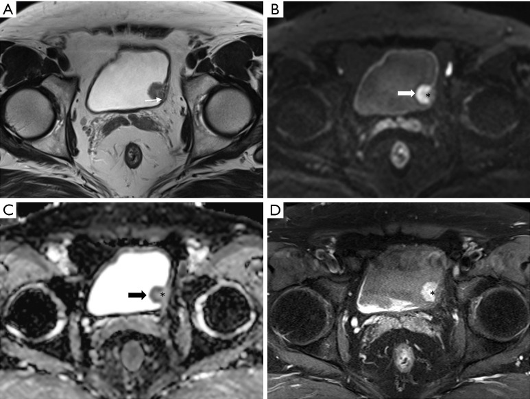
Figure 9.
Urothelial carcinoma (stage T1) in a 60-year-old man. Axial (A) T2-weighted images showing a tumor in the left bladder wall. The muscle layer with a T2 hypointensity line is suspicious of focal disruption because of the presence of tumors with T2 intermediate signal intensity (thin arrow). In the high b value (b =800) diffusion-weighted image (B) and apparent diffusion coefficient map (C), the peripheral region of the tumor shows marked diffusion restriction (thick arrow). However, diffusion restriction is not seen at the base of the tumor, including the stalk (asterisk). Additionally, disruption of the muscle layer with intermediate signal intensity is not observed even in high b value images. An axial three-dimensional T1-weighted spoiled gradient echo image (D) obtained 300 seconds after contrast agent administration shows bright delayed enhancement of the tumor stalk (asterisk). This case was preoperatively classified as vesicle imaging-reporting and data system grade 2. The patient underwent transurethral resection of the bladder tumor. Appropriate tumor specimens with muscle layer were obtained and it was diagnosed as high-grade stage T1 cancer with invasion only up to the subepithelial connective tissue.

