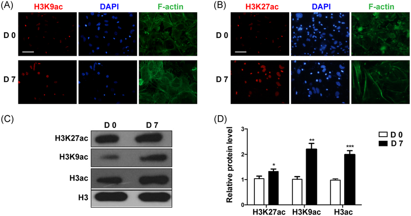
FIGURE 3.
The expression of acetylated H3K9 and H3K27 during odontoblast differentiation of the mDPCs. mDPCs were cultured with odontoblastic differentiation medium for 7 days. Immunofluorescence was performed to detect the expression levels of acetylated H3K9 (A) and H3K27 (B); the nucleus was labeled with DAPI. The protein levels of acetylated H3K9 and H3K27 (C, D) were detected. The D0 group was set as the control. All data were presented as mean ± standard deviation (SD) and were based on three independent experiments. DAPI, 4’,6-diamidino-2-phenylindole; mDPCs, mouse dental papilla mesenchymal cells. *P < .05, **P < .01, ***P < .001

