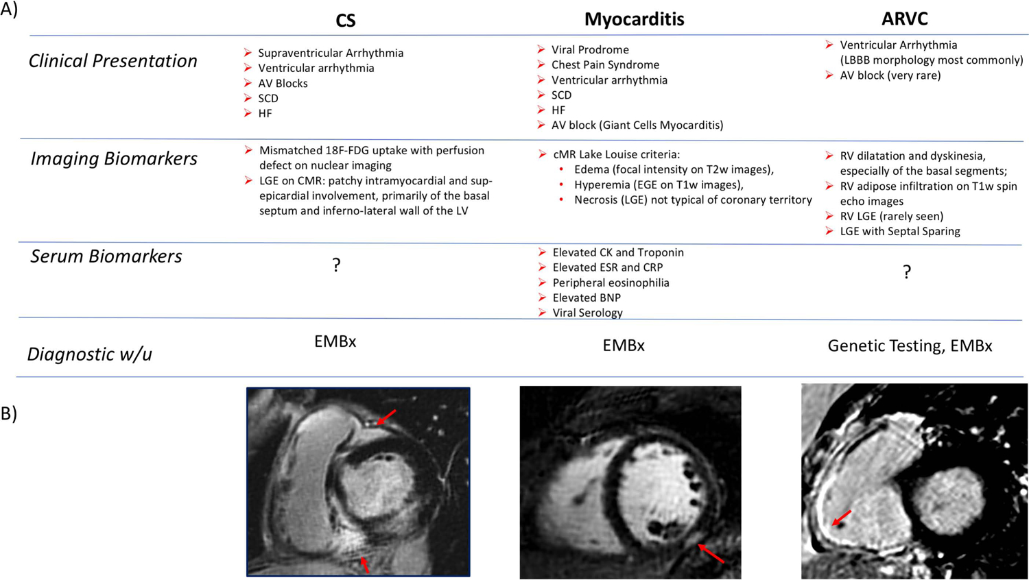
Figure 4: Diagnosis of ICS.

(A) Clinical, Imaging and Serological Biomarkers that might aid in the DDx of CS. (B) Short axis views of RV and LV showing classical patterns of LGE involving the 1) RV insertion point, basal septum as well as lateral wall in CS, 2) the sub-epicardium in myocarditis and 3) the RV free-wall in ARVC.
