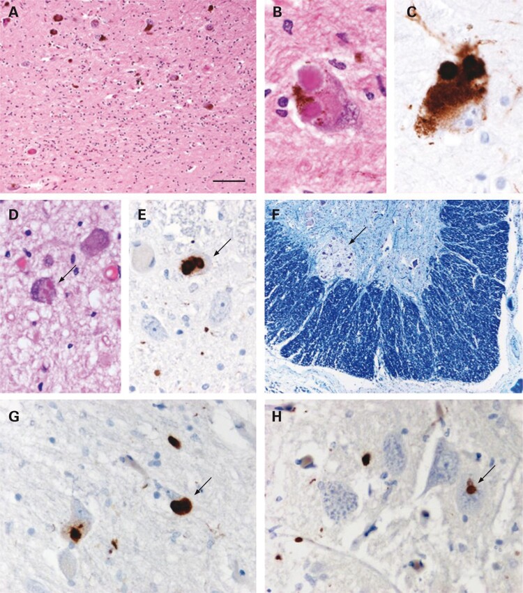
Figure 2.
Examination of the substantia nigra showed moderate loss of pigmented neurons with gliosis (A). Typical Lewy bodies were identified in residual neurons (B) and were confirmed using α-synuclein immunohistochemistry (C). Lewy bodies were also found in other brain stem structures, including the Xth nerve nucleus (arrow in D). Immunohistochemical staining for α-synuclein also revealed small numbers of Lewy bodies in neurons of the intermediolateral column in the thoracic spinal cord (arrow in E). In the sacral cord, the Onuf’s nucleus (ON) was identified (arrow in F) and, at this level, Lewy neurites and Lewy bodies could be identified in the gray matter (arrow in G indicates a Lewy body). In the ON, there were Lewy neurites and also a single probable Lewy body (arrow in H). A, B and D: Haematoxylin and eosin; F: Luxol fast blue and cresyl violet; C, E, G and H: α-synuclein immunohistochemistry, chromogen: diaminobenzidine. Bar in A represents 260 μm in F; 100 μm in A; 25 μm in D, E, G and H; and 17 μm in B and C. Reproduced with permission from O’Sullivan et al.31

