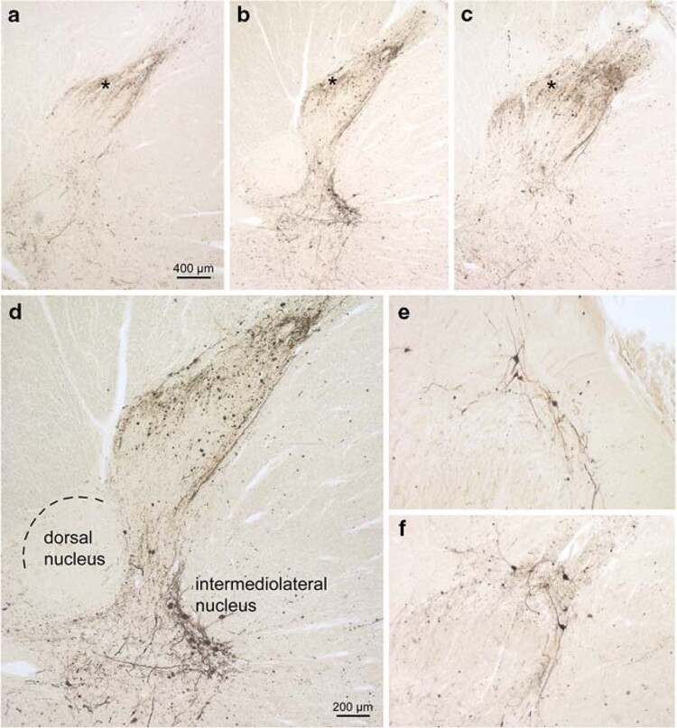
Figure 3.
PD-related involvement of the spinal cord. a Seventh cervical segment. b Twelfth thoracic segment. c Third lumbar segment. Note the increase caudally in the density of the LN network in lamina I (asterisks) and close to it. d Detailed micrograph of b showing involvement of the intermediolateral nucleus. Several multipolar preganglionic sympathetic relay neurons are Wlled with _-synuclein aggregates. Note that the dorsal nucleus (Clarke’s column) is virtually uninvolved. a–d orginate from a 76-year-old male at stage 4 (case 2). e, f Both sections show the multipolar relay neurons in lamina I as being exclusively aVected. In contrast, the nerve cells in the subjacent layer II remain intact. e Originates from the seventh cervical spinal cord segment of case 6, f from the tenth thoracic spinal cord segment of case 3. Syn-1 (Transduction Laboratories) immunoreactions in 100 _m polyethylene glycol-embedded tissue sections. Scale bar in a is valid for b and c. Scale bar in d applies to e and f. Reproduced with permission from Braak et al.36

