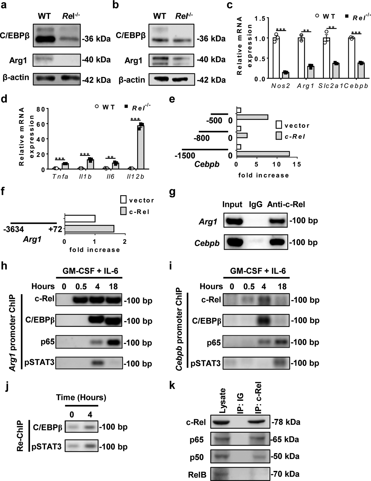
Fig. 4. c-Rel activates MDSC signature genes including Cebpb and Arg1.

a, b, C/EBPβ and Arg1 protein expression in MDSCs from WT and Rel−/− mice that had similar size of B16F10 tumors, or bone marrow-derived MDSCs as determined by Western blot.
c, d, Relative mRNA levels of downregulated (c) and upregulated (d) genes in bone marrow-derived Rel−/− MDSCs as compared to WT MDSCs, determined by real-time RT-PCR (n=3 biologically independent cultures/group; **, P<0.01; ***, P<0.001)
e, f, The effect of Rel gene on Cebpb (e) and Arg1 (f) gene promoter regions as determined in the luciferase reporter assay. The results are representative of two independent experiments.
g, c-Rel binding to the Cebpb and Arg1 gene promoter regions in bone marrow-derived MDSCs as determined by chromatin immunoprecipitation (ChIP). Control IgG and input DNA are shown as controls.
h-i, The binding kinetics of c-Rel, C/EBPβ, p65, and pSTAT3 to the Arg1 (h) and Cebpb (i) gene promoters in cultured bone marrow cells treated with GM-CSF and IL-6 for up to 18 hours.
j, The presence of the enhanceosome complex at the Cebpb promoter in cultured bone marrow cells treated with (4 hours) or without GM-CSF and IL-6, as determined by first ChIP with anti-c-Rel and Re-ChIP with anti-C/EBPβ and anti-pSTAT3.
k, Co-immunoprecipitation (co-IP) of c-Rel with the indicated proteins (marked on the right), of bone marrow-derived MDSCs from WT mice.
Statistical significance was determined by two-tailed unpaired t-test (c-f). Data are presented as means ± s.e.m (c-f).
