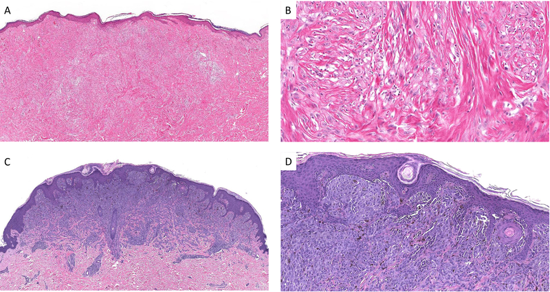
Figure 1.
An example of a desmoplastic Spitz nevus with 11 p gain (case #4). The lesion is densely fibrotic with a pink appearance (A) Amelanotic spindle and epithelioid melanocytes are dispersed in a fibrotic stroma at low cell density (B). Example of a Spitz nevus with 11 p gain and only minimal sclerosis (case #2). There is a sharply circumscribed compound melanocytic proliferation with evidence of maturation (C). Nests of spindle and epithelioid melanocytes with focal melanin pigment are seen in the superficial aspect of the lesion (D).

