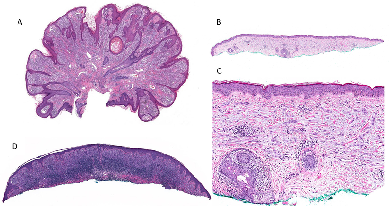
Figure 2.
Low-power image showing case #3 with a markedly papillomatous surface with hyperplastic epidermis, high tumor cellularity, and sparse perivascular lymphocytic infiltrate (A). Case #7 is an example of a pauci-cellular tumor with flat surface, plaque-like distribution of melanocytes in the dermis, and no associated epidermal changes (B) composed of epithelioid and spindle cells without significant stromal desmoplasia (C). Case #23 showing a dense associated band-like lymphocytic infiltrate (D).

