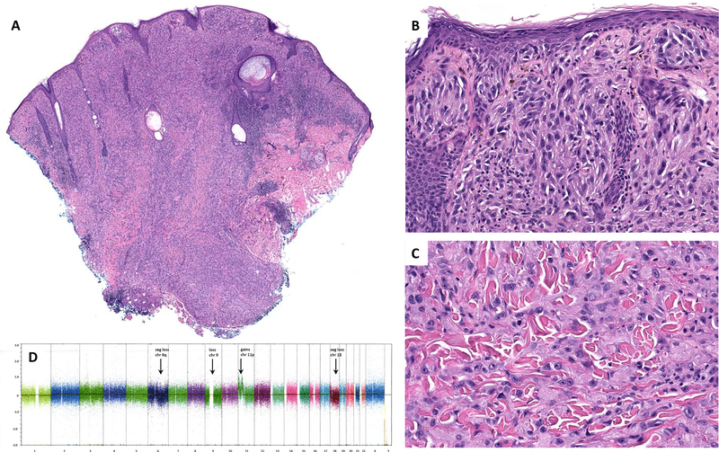
Figure 3.
Low-power image of case #31 showing a raised surface, sheet-like growth pattern of tumor cells and nodular/bulbous pushing borders that extend into superficial subcutis (A). Nests of plump spindle and epithelioid melanocytes are present at the dermoepidermal junction and in the superficial dermis (B). Interstitial pattern of predominantly epithelioid melanocytes in the deep dermis (C). SNP array (D) showed gains in 11p and low level segmental loss in 6q, loss of chromosomes 9, segmental loss in chromosome 18, and CN-LOH of a segment of chromosome 19. seg = segmental; chr = chromosome

