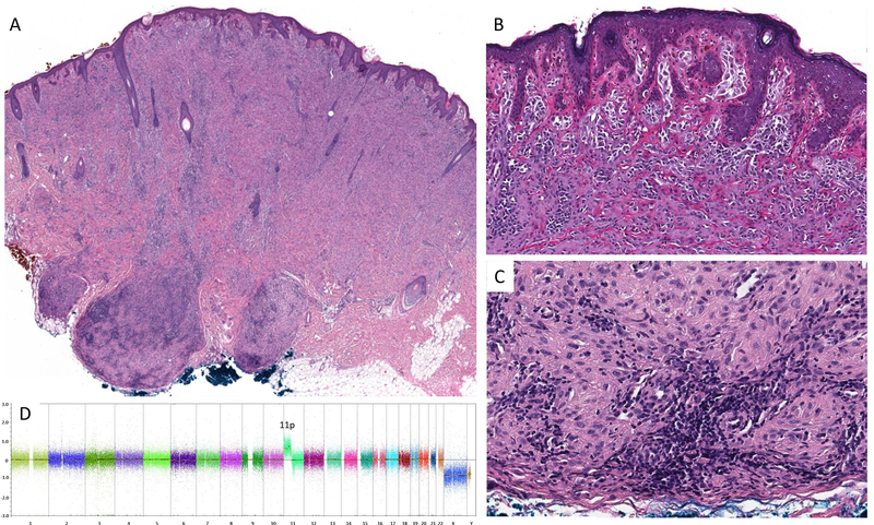
Figure 4.
Silhouette of the tumor from the cheek of a 3 year-old boy (Case #9). The tumor shows two small bulbous projections at its base (A). Superficial portion of nests of plump spindle and epithelioid cells (B). Aggregates of epithelioid melanocytes in the deep bulbous portion of the tumor are associated with lymphocytes (C). No cytogenetic aberrations other than an isolated gain of 11p were found in this tumor (D).

