
Figure 1.
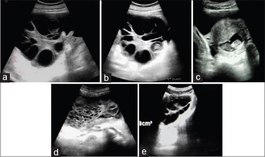
(a and b) Case 1: Transverse sections of enlarged right and left ovaries with thick vascularized echogenic stroma containing multiple peripherally placed thin-walled cysts some of which show internal echoes. (c) Case 1: Single viable intrauterine fetus with a normal anteriorly sited placenta. (d) Case 2: Bulky uterus containing a hyperechoic structure with multiple small-sized cystic spaces within its cavity. (e) Case 2: Bulky right ovary with peripherally placed thin-walled anechoic cysts and a thickened echogenic stroma giving a spoke wheel appearance