Figure 10.
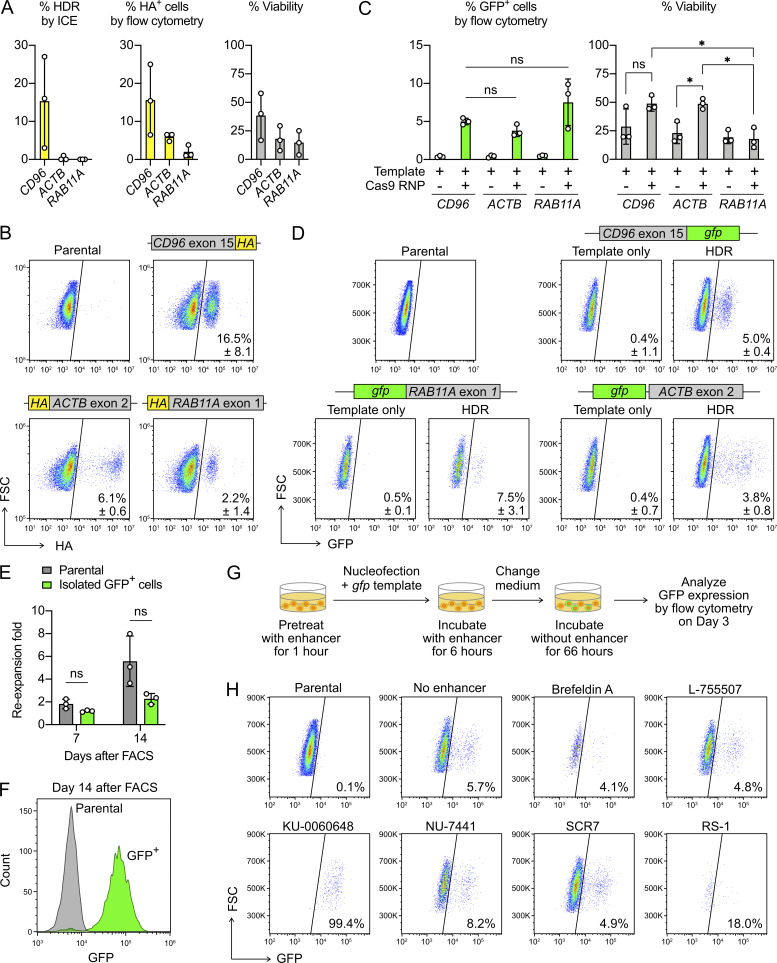
Nonviral gene KI is feasible in primary NK cells. (A) HA tag KI efficiencies at the CD96, ACTB, and RAB11A loci were determined by ICE and flow cytometry. The percent viability was analyzed by Precision beads assay. (B) Representative flow cytometry plots with the mean KI rates ± SD. The positions of HA tag KI are indicated. (C) The gfp KI efficiencies as determined by flow cytometry. (D) Representative flow cytometry plots with the mean KI rates ± SD. The positions of gfp KI are indicated. (E) Reexpansion rate of the FACS-isolated GFP+ cells that carried the gfp KI at the CD96 locus. (F) Representative flow cytometry histogram shows the expression of GFP after 14 d of reexpansion. (G) Screening of HDR enhancers to increase the gfp KI efficiency at the CD96 locus. (H) Flow cytometry plots show the percentage of GFP+ cells from different enhancer treatments. Cell density was normalized by Precision beads count to reveal reduction in the cell number in some treatments. Data are shown as mean ± SD of three donors (n = 3), except that HDR enhancers were tested in one donor (n = 1). Two-tailed Welch’s unequal variances t test was used to test for statistical significance. *, P ≤ 0.05. FSC, forward scatter; ns, not significant.

