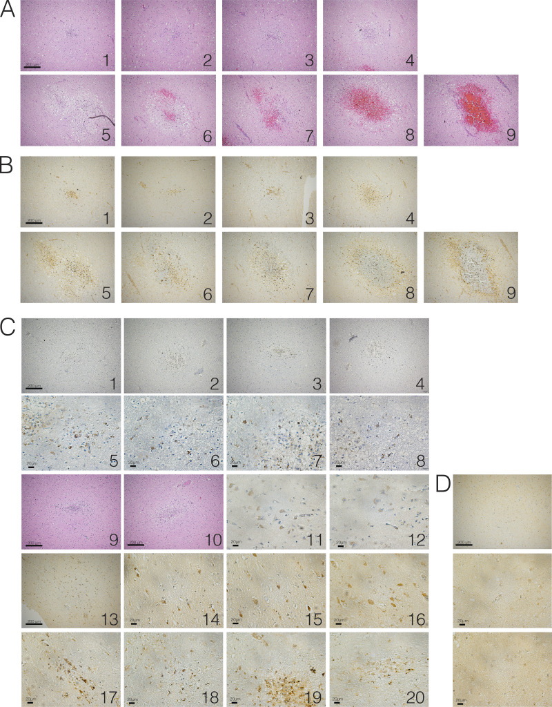
Figure S5.
Evidence of SARS-CoV-2 neuroinvasion–associated ischemic infarcts. FFPE sections of brain tissue from COVID-19 patients were stained using H&E or anti–SARS-CoV-2-spike antibody. (A) H&E images of ischemic infarcts at different stages (1, earliest, to 9, latest). (B) SARS-CoV-2–stained images of ischemic infarcts at different stages (1, earliest, to 9, latest). Each number corresponds to H&E image in A. (C) Images of SARS-CoV-2–positive regions in brains of COVID-19 patients. (D) Example image from control patient brain. Scale bar = 200 µm for zoomed-out images and 20 µm for zoomed-in images.

