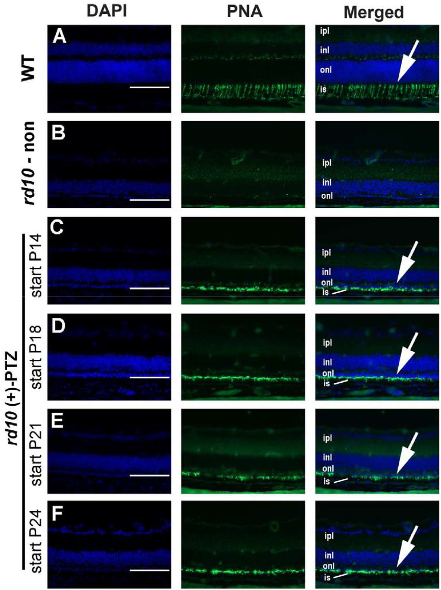
Figure 5. Evaluation of cone photoreceptor labeling.

Following the in vivo studies, one eye per mouse was processed for cryosectioning and immunodetection of FITC-conjugated peanut agglutinin (PNA), a green fluorescent marker of cone PRCs. Representative photomicrographs are shown for (A) WT, (B) non-treated rd10 mouse, or rd10 mice administered (+)-PTZ starting on (C) P14, (D) P18, (E) P21, (F) P24. (Arrows in A, C-F point to green fluorescing cone inner segments.) Abbreviations: ipl, inner plexiform layer; inl, inner nuclear layer; onl, outer nuclear layer; is, inner segments. Calibration bar = 100μm.
