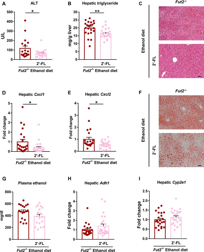
Figure 4. Restoration of α1-2-fucosylated glycans in the intestine attenuates ethanol-induced liver disease in Fut2 deficient mice.
Fut2−/− mice were assigned to 2’-fucosyllactose (2’-FL) treated group and control group, and fed with chronic-binge ethanol diet (NIAAA model). In the 2’-FL treated group, 2’-FL (2mg/mL) was supplemented continuously in the ethanol diet. The experimental diet and 2’-FL treatment lasted for 15 days. (A) Plasma alanine aminotransferase (ALT). (B) Hepatic triglyceride levels. (C) Representative images of H&E-stained liver tissue. (D) Hepatic Cxcl1 mRNA. (E) Hepatic Cxcl2 mRNA. (F) Representative images of Oil Red O-stained liver tissue. (G) Plasma ethanol. (H) Hepatic expression of Adh1 mRNA. (I) Hepatic expression of Cyp2e1 mRNA. Data represent mean ± SEM; * and ** indicate P<0.05 and P<0.01, respectively. Scale bar = 50μm. Experiments performed in n=18–24 per group. For the H&E and Oil Red O staining, n=10 per group.

