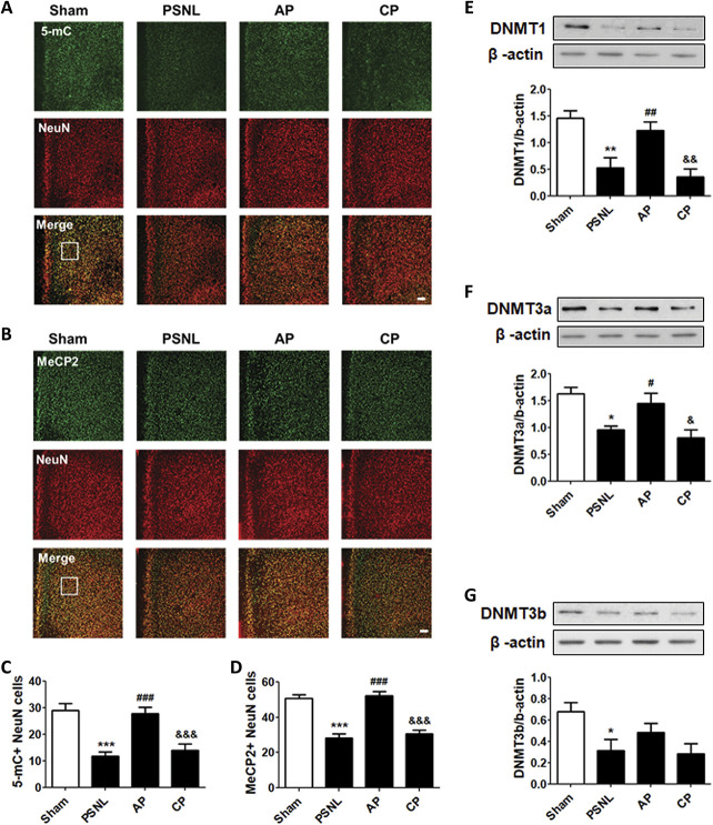
Figure 7.
Effects of acupuncture on the expression levels of 5-mC, MeCP2, and DNMTs in the PFC. Histological examinations of the PFC showing expression of 5-mC (green) (A–C) and MeCP2 (green) (B–D) with NeuN (red) after acupuncture administration (AP or CP) for 6 months (3 days/week) in a PSNL-induced neuropathic pain model (A and B); this representative image shows 5-mC-positive neuron cells (C) and MeCP2-positive neuron cells (D) in the PFC region (n = 6/group). Scale bar: 100 μm. ***P < 0.01 vs sham group, ###P < 0.001 vs PSNL group, and ###P < 0.001 vs PSNL group. The results showed the changes in protein expression levels of DNMT1 (E), DNMT3a (F), and DNMT3b (G) in the PFC after administration of AP or CP for 6 months (n = 4/group, *P < 0.05, **P < 0.01 vs sham group, #P < 0.05, ##P < 0.01 vs PSNL group, and &P < 0.05, &&P < 0.01 vs AP group). The results were analyzed using one-way ANOVA followed by the Newman–Keuls post hoc test. All data are expressed as the mean ± SEM. 5-mC, 5-methylcytosine; ANOVA, analysis of variance; AP, acupuncture points; CP, control point; CpG, cytosine-phospho-guanine; DNMT, DNA methyltransferase; MeCP2, methyl-CpG binding protein 2; PFC, prefrontal cortex.

