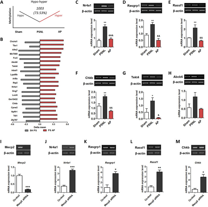
Figure 8.
Effects of acupuncture on the methylation levels of cell death-related genes in the PFC. The graphs showing the genes of the hypo-hyper methylation pattern (A) and top 20 in the hypo-hyper methylated genes (B) in the PFC. The graphs show the changes in the mRNA expression of Nr4a1, Rasgrp1, Rassf1, Chkb, Tekt4, and Abcb6 (C-H) in the PFC after administration of AP or CP for 6 months. The graphs show changes in the mRNA expression of Mecp2, Dnmt1, Dnmt3a, and Dnmt3b at 3 days after treatment with Mecp2 siRNA in the neuronal cell of the mouse frontal cortex (I–M). n = 3/group, *P < 0.05, **P < 0.01 vs sham group, #P < 0.05, ##P < 0.01, ###P < 0.001 vs PSNL group. All data were analyzed using one-way ANOVA followed by the Newman–Keuls post hoc test. All data are shown as mean ± SEM. ANOVA, analysis of variance; AP, acupuncture points; CP, control point; PFC, prefrontal cortex; PSNL, partial sciatic nerve ligation; siRNA, small interfering RNA.

