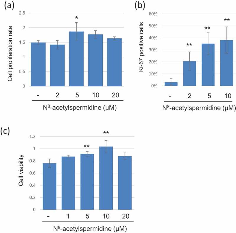
Figure 5.

Induction of cell growth and chemoresistance to colon cancer cells by N8-acetylspermidine. (a) HCT116 cells were cultured with FBS-free culture medium containing N8-acetylspermidine, and then, cell viability was measured using CCK8 kit at 24 h and 72 h. The cell proliferation rate was calculated as the cell viability at 72h/cell viability at 24 h. (b) HCT116 cells were cultured with FBS-free culture medium containing N8-acetylspermidine for 72h, and the cells were immunocytochemistry stained with proliferation marker protein Ki-67 and DAPI. The percentage of Ki-67 positive cells was counted from randomly taken 10 images. (c) HCT116 cells were cultured with culture medium containing 20 µM oxaliplatin and N8-acetylspermidine. After 48 h, the cell viability was measured using CCK8. Data represent the mean (± standard deviation, SD) of three independent experiments, each performed in triplicate. Error bars indicate SDs. Asterisks indicate the statistical significance of each concentration of N8-acetylspermidine versus distilled water (* P<0.05, ** P<0.01; Tukey’s test)
