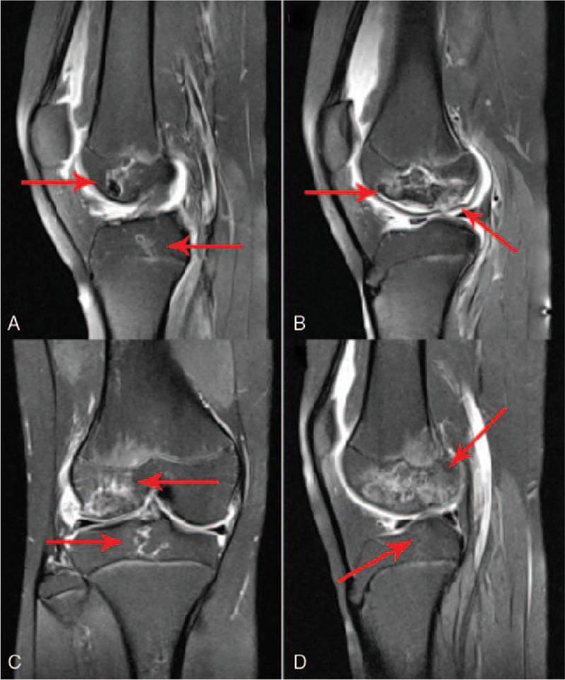
Figure 1.

Bilateral knee joint magnetic resonance imaging appearance of the systemic lupus erythematosus patient with osteonecrosis (A) and (B) Sagittal magnetic resonance imagings of left knee joint. (C) and (D) Sagittal magnetic resonance imaging of right knee joint. The figure demonstrated irregular bone destruction and bone hyperplasia lesions on bilateral distal femur and proximal tibia, presenting geographic alterations.
