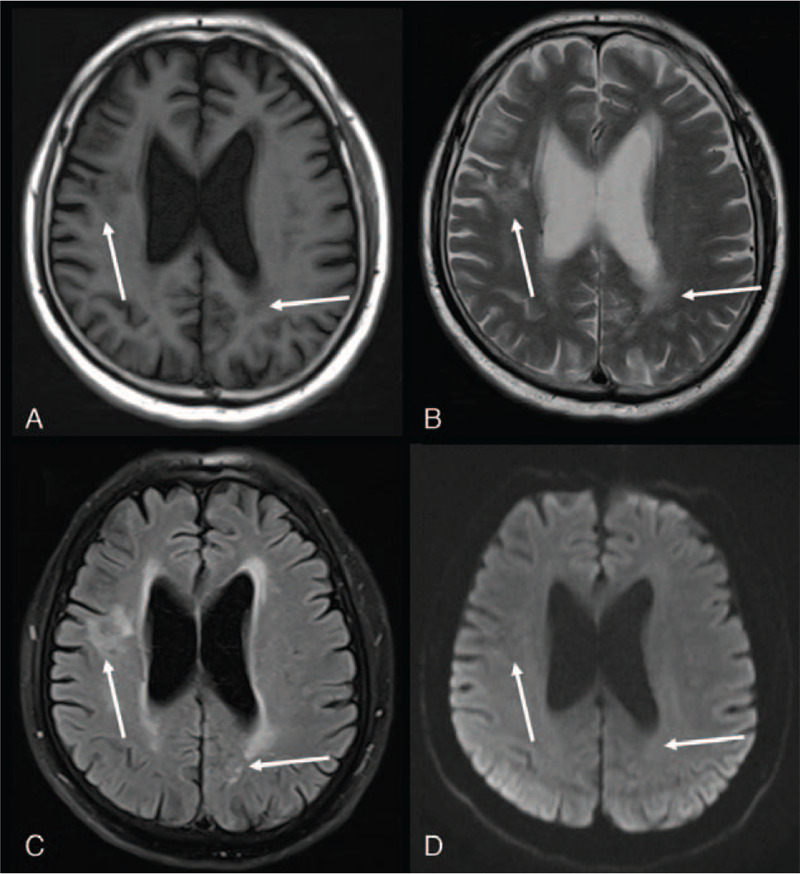
Figure 3.

Reexamination of the brain MRI one and a half years later showed resolution of lesions with small softened foci and glial hyperplasia. A, Axial T1-weighted image. B, Axial T2-weighted image. C, Axial T2 Flair-weighted image. D, Diffusion-weighted image (b = 800). MRI = magnetic resonance imaging.
