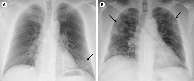
Fig 1.
Example radiographs of (A) low-risk CXR and (B) high-risk CXR. (A) Low-risk CXR showing unilateral opacity (demarcated by black arrow) in the left lower zone. This patient’s symptoms resolved after 3 days and he did not require supplemental oxygen throughout his admission. (B) High-risk CXR showing bilateral, multifocal opacities involving the upper zones (demarcated by black arrows). This patient developed hypoxia requiring supplemental oxygen during his admission.

