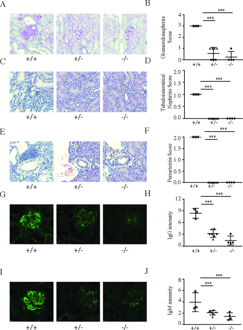
Fig 5. Lack of SLC15A4 protects mice from developing spontaneous lupus nephritis in NZB/W F1 mice.

NZB/W F1 slc15a4+/+, slc15a4+/-, and slc15a4-/- mice were euthanized at the age of 36 weeks. H&E and PAS staining was carried out on formalin fixed tissues to assess renal disease severity by comparing severity scores of glomerulonephritis (A, B), tubulointerstitial nephritis (C, D) and periarteritis (E, F). Immune-complex deposition in kidney was assessed in frozen tissue sections by staining for IgG (G,H) and IgM (I,J) and quantified using FITC/AF488 integrated intensity in renal cortex. Examples (A, C, E, G, I) and quantification over several samples (B, D, F, H, J) is shown. Data is plotted with group means ± SD; individual data points represent results from two kidney sections on one slide from an individual mouse. *p < 0.05, **p<0.005, ***p<0.0005.
