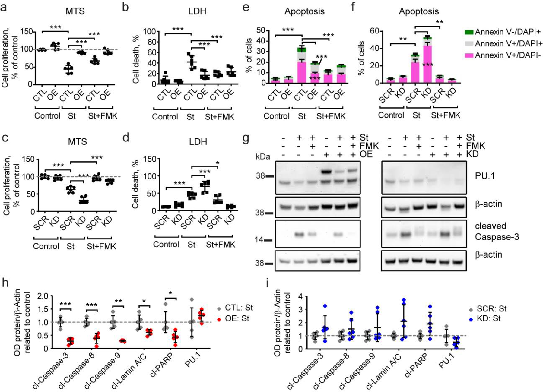
Figure 3. Stable PU.1 overexpression decreases and knock-down increases apoptotic cell death in BV2 microglial cells.

(a, b) PU.1 overexpression leads to increased resistance of BV2 cells to staurosporine treatment, while (c, d) PU.1 knock-down cells are more sensitive to staurosporine treatment measured by MTS (a, c) and LDH (b, d) assays. (e) PU.1 overexpression decreases the number of early apoptotic and necrotic cells after staurosporine treatment. (f) PU.1 knock-down increases the number of early apoptotic cells after staurosporine treatment. (g) Activation of caspase-3 was reduced in PU.1 overexpressing cells and unaffected in PU.1 knock-down cells after staurosporine treatment, representative western blot image. (h, i) Quantification of apoptosis-dependent activation of proteins based on western blot as shown in (g) and Supplementary Fig. S5. Values shown are mean ± SD of (a-d) n = 3 independent experiments with two technical replicates each or (g–i) n = 4–5 independent experiments with one technical replicate. * P < 0.05, ** P < 0.01, *** P < 0.001. (a–d) One-way ANOVA with Bonferroni’s post-hoc multiple comparisons, (h–i) unpaired t test with Welch’s correction.
