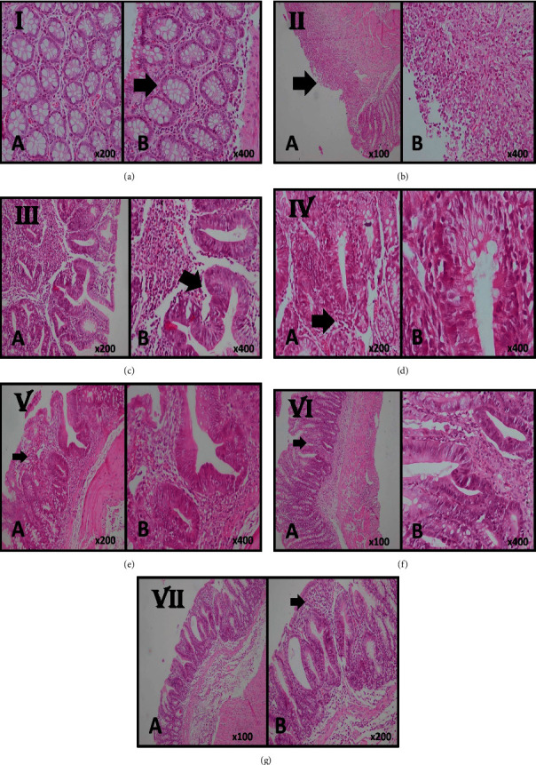
Figure 2.

Histological sections of colonic mucosa in case and control groups. (a) Normal mucosa with the intact epithelial surface as well as normal colonic crypts (arrow) in the sham control group (A, B H & E stain, ×200, ×400). (b) In the negative control group, the slide shows TNBS-induced colitis with severe destruction with surface ulceration (arrow) and severe inflammatory cell infiltration representative of chronic colitis with severe activity, (A, B H & E stain, ×100, ×400). (c) Colonic mucosa of the cases who received 200 mg/kg QB extract orally shows moderate infiltration of inflammatory cells in lamina propria and mild crypt destruction and loss of goblet cells and cryptitis (arrow) representative of chronic colitis with mild activity (A, B H & E stain, ×200, ×400). (d) Colonic mucosa of the cases who received 400 mg/kg QB extract orally shows mild infiltration of inflammatory cells in lamina propria (arrow) and no crypt destruction and with few crypts with loss of goblet cells (A, B H & E stain, ×200, ×400). (e) Colonic mucosa of the cases who received 200 mg/kg QB extract rectally shows mild infiltration of inflammatory cells in lamina propria (arrow) and no crypt destruction and with few crypts with loss of goblet cells (A, B H & E stain, ×200, ×400). (f) Colonic mucosa of the cases who received 400 mg/kg QB extract rectally shows near-normal colonic mucosa with few inflammatory cell infiltrates in lamina propria (arrow) and focal crypts with loss of goblet cells (A, B H & E stain, ×100, ×400). (g) Colonic mucosa of the treated rats with 500 mg/kg of sulfasalazine shows near-normal colonic mucosa with focal crypts with loss of goblet cells with few inflammatory cell infiltrates in lamina propria (arrow) (A, B H & E stain, ×100, ×200).
