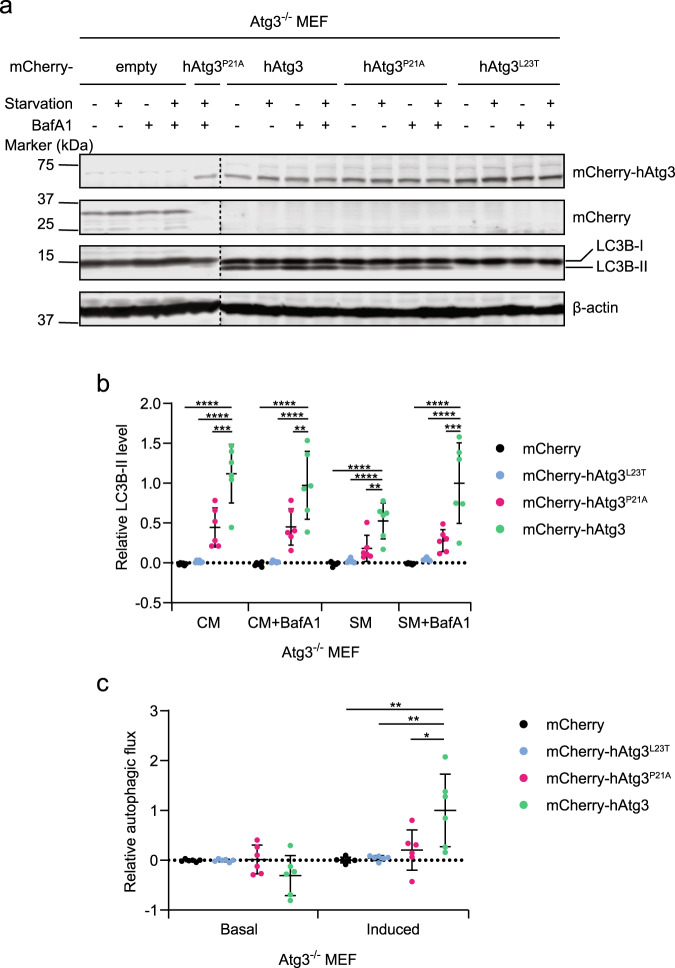
Fig. 4. hAtg3P21A and hAtg3L23T are impaired in inducing LC3B lipidation and autophagic flux in vivo.
Atg3 knockout (Atg3−/−) mouse embryonic fibroblasts (MEFs) stably expressing mCherry, mCherry-hAtg3, mCherry-hAtg3P21A, or mCherry-hAtg3L23T were cultured in complete media (CM) and starvation media (SM) with or without bafilomycin A1 (BafA1) for 3 h and subjected to immunoblotting with the indicated antibodies. a Representative immunoblot (n = 6 blots). b Quantitative analysis of the relative LC3B–II level (n = 6 blots). c Relative basal and starvation-induced autophagic flux (n = 6 blots). Statistical analysis was performed using one-way ANOVA test followed by Dunnett’s multiple comparisons test. Data are presented as mean ± SD. P values: ****P < 0.0001 (b), ***P = 0.0001 and 0.0003 (left to right in b), **P = 0.0037 and 0.0011 (left to right in b) and 0.0014 and 0.0021 (left to right in c), and *P = 0.0097 (c).

