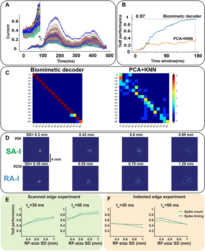
Figure 8.
The performance of the tactile spiking neural network when stimulus location is changed on the skin. (A) Input currents of cortical neurons: blue traces show those neurons that are tuned to the presented input stimulus, each of them responds to a stimulus which is applied at a different skin region. The magnified part indicates the transient phase of indentation. Using the cortical neuron currents, it is found that indented edges can be detected after (time delay to convey contact information to the cortex). (B) The superior performance of the biomimetic decoder in recognizing edge stimuli indented at a different position on the simulated skin. (C) Confusion matrix for two classifiers, the biomimetic and the KNN classifiers. (D) Simulated receptive fields of SA-I and RA-I for 4 different scales (SD (mm)) based on the Gaussian distribution of the innervated mechanoreceptors. (E) Exploring the effect of afferent receptive field size on the temporal and spatial coding of the cortical neurons. Network Performance for spike counting (green) and spike timing (blue) when edge stimulus is scanned across the skin for (left) and (right) after contact. (F) Network Performance for spike counting (green) and spike timing (blue) for (left) and (right) after contact.

