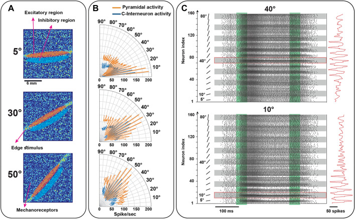
Figure 9.
The cortical neuron receptive fields with excitatory and inhibitory sub-regions on the skin. (A, B) Receptive fields of cortical neurons are located at different positions across the skin and neurons selectively respond to the orientation stimulus. Activation of excitation areas on the skin increases the cortical firing, on the other hand, activation of inhibitory areas leads to a decrease in the spontaneous firing. (C). The firing response of 160 orientation-sensitive neurons in area 3b to 10° and 40° indented edge stimuli. Each group of the ten neurons is sensitive to one orientation which location of their receptive fields on the skin is different. The green areas show the onset and end phases of indentation.

