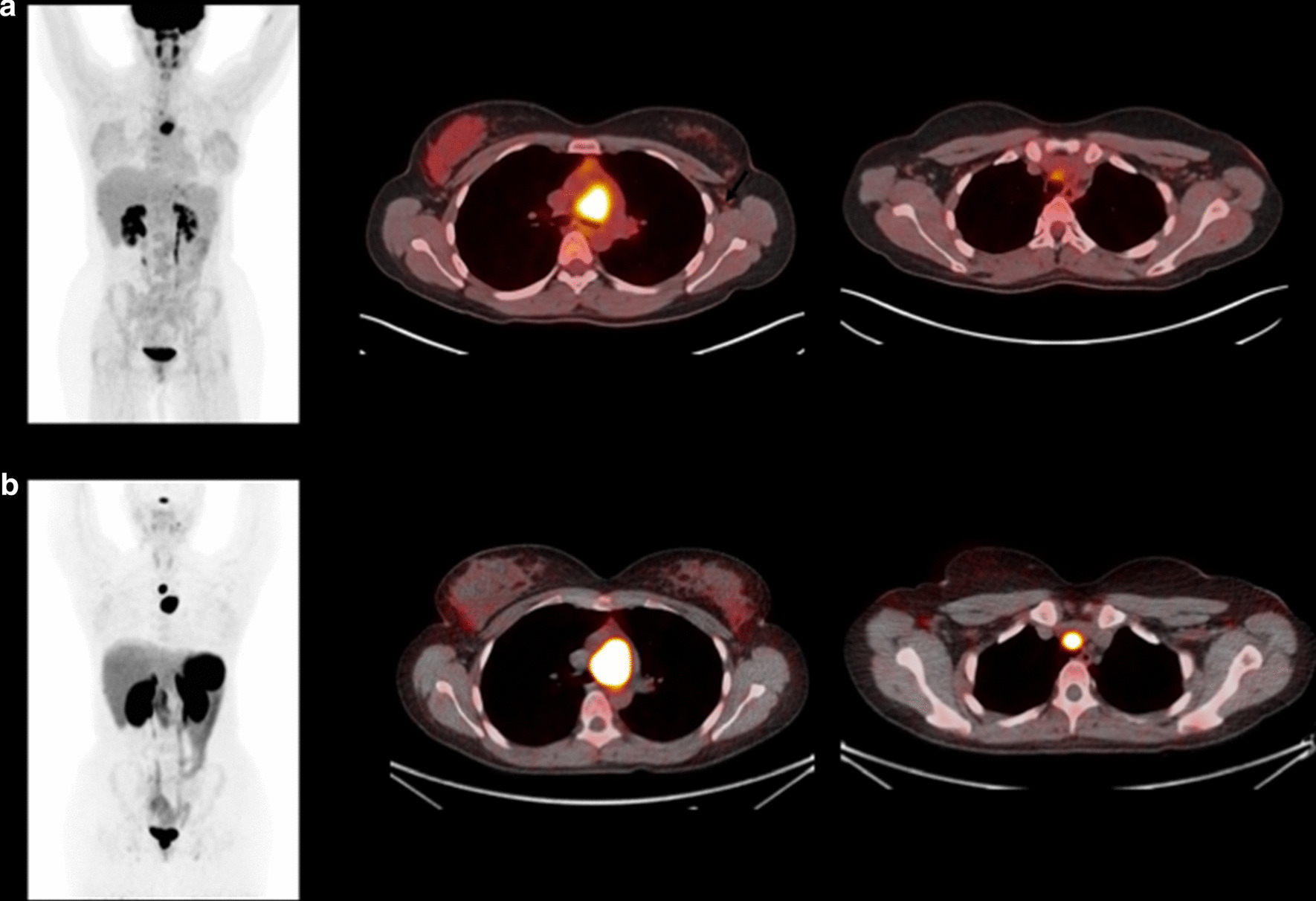
Fig. 4.

Case 012. Panel A, [18F]FDG PET/CT MIP image and fused transaxial views showing a highly avid mediastinal mass and low-level uptake of an adjacent lymph node (C). This imaging was performed as part of surveillance for disease recurrence given a background of a resected wild-type GIST, and the mediastinal mass was a presumed recurrence of this earlier tumour. Panel B, 68 Ga-DOTATATE PET/CT, same views as above. Very intense uptake in the mediastinal mass (SUVmax 45) and adjacent lymph node (SUVmax 40). This patient had normal plasma metanephrine and 3-methoxytyramine values on biochemical testing. Biopsy confirmed a non-secreting mediastinal dSDH paraganglioma case 012
