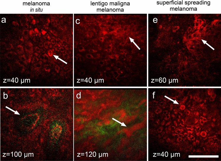
Figure 4.
MPM features of melanoma in dermoscopic equivocal group. In melanoma in situ, large pagetoid cells in spinous layer (arrow, a) and non-edged papillae (arrow, b) are marked. In lentigo maligna melanoma, large pleomorphic cells in spinous layer (arrow, c) and melanophages below dermal–epidermal junction (arrow, d) are marked. In superficial spreading melanoma, nest of atypical cells (arrow, e) and round pagetoid cells (arrow, f) in the epidermis are marked. Autofluorescence from the cells (red) and SHG signal from collagen (green) are shown. Imaging depth is marked on the images. Scale bar is 100 µm, applicable to all images.

