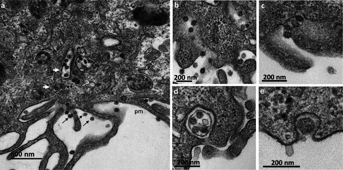
Fig. 5.
Release of the SARS-CoV-2 virions by exocytosis from the Vero cells at 10 h post-infection. (a) Virions (thin black arrows) were frequently observed at the plasma membrane (pm). Numerous virus-carrying vesicles (white arrows) presumably in transit to the plasma membrane were also visualized. (b–e) These virus-carrying vesicles fused with the plasma membrane to release their contents into the extracellular space by exocytosis. Similar ultrastructural events were observed at 12 hpi

