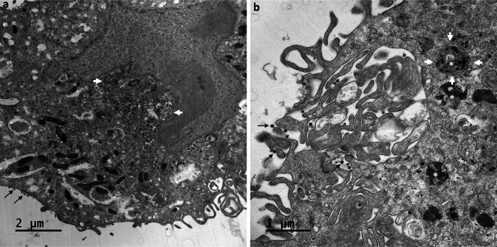
Fig. 6.
Intracellular accumulation of SARS-CoV-2 particles in large vacuoles within Vero cells at 24 hpi. (a,b) Viruses were still detected at the cell surface (thin black arrows), but large numbers of viral particles were observed accumulated in very large intracellular vacuoles (white arrows) of various sizes, predominantly located in the perinuclear region. At this time point, an accumulation of myelin-like membrane whorls or autophagic-like packaged membranes (white asterisk in b) was also observed in the cells, separated from or associated with the viral particles

