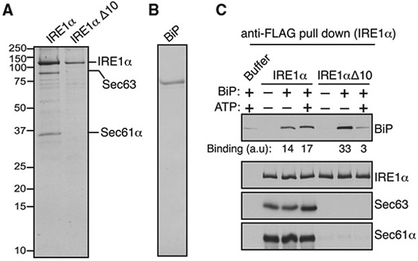
Figure 5. Biochemical Reconstitution of Sec61/Sec63-Mediated BiP Binding to IRE1α.

(A) A Coomassie-blue-stained gel showing the purified IRE1α/Sec61/Sec63 complex or IRE1α Δ10 from HEK293 stably expressing 2xStrep-IRE1α-FLAG or 2xStrep-IRE1α ΔD10-FLAG.
(B) A Coomassie-blue-stained gel showing purified His-BiP from E. coli.
(C) The purified IRE1α/Sec61/Sec63 complex or IRE1αΔ10 was bound to anti-FLAG beads and incubated with or without BiP in the presence or absence of ATP as shown. After incubation, IRE1α-bound anti-FLAG beads were washed and eluted with sample buffer. A negative control, the reaction was performed by incubating empty anti-FLAG beads with the buffer, BiP, and ATP. The samples were analyzed by immunoblotting for the indicated antigens. BiP bands were quantified and presented as arbitrary units (a.u) after subtracting the buffer background.
See also Figure S6.
