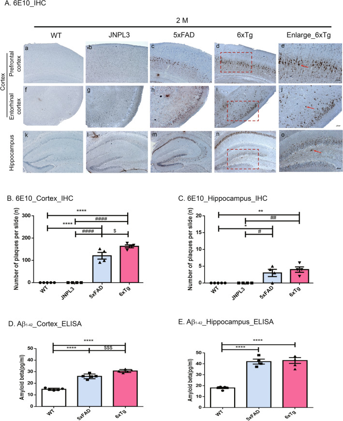
Figure 2.
Age-dependent increase of Aβ plaques and amount of Aβ in the brains of 6xTg mice. (A) Immunohistological staining of amyloid plaques and tau phosphorylation with anti-Aß (6E10) in the cortex [prefrontal cortex (a–d) and entorhinal cortex (f–i)] and hippocampal dentate gyrus (k–n) of 2-month-old 6xTg mice and their age- and gender-matched WT, 5xFAD, and 6xTg littermates. Scale bar 200 μm. The red box contains enlarged images of cortical and hippocampal regions of 6xTg mice (e, j, o; Scale bar 100 μm). Red arrow-shaped 6E10-positive neurons were counted. (B, C) In the 6xTg mice, the number of plaques was significantly increased compared with WT mice, but Aß deposition showed no significant difference compared with the 5xFAD mice, but showed a tendency to increase in the hippocampus. (D, E) Aß proteins from cortical and hippocampal brain lysates of mice were subjected to ELISA assay and levels of Aß were examined. All data are given as means ± SEM (n = 5 mice per group). Statistical analyses were performed by one-way ANOVA followed by the Tukey's multiple comparisons test. ****p < 0.0001, ***p < 0.001, **p < 0.01 vs, *p < 0.05 vs. WT; ####p < 0.0001, ###p < 0.001, ##p < 0.01, #p < 0.05 vs. JNPL3; $$$p < 0.001 vs, $p < 0.05 vs. 5xFAD.

