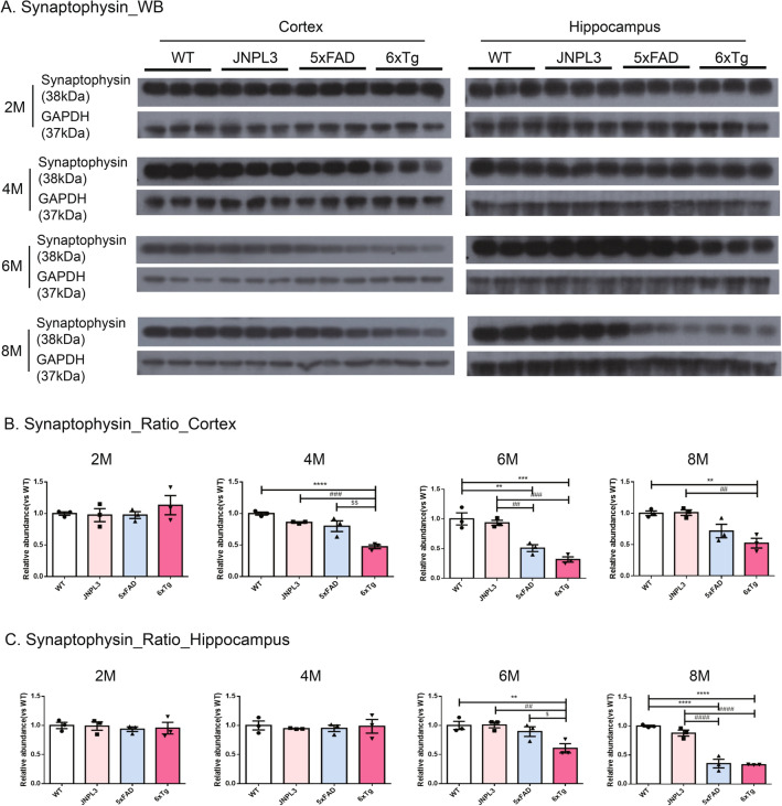
Figure 5.
Synaptophysin levels in the cortical and hippocampal area of 6xTg mice brains. Presynaptic receptor proteins (Synaptophysin, 38 kDa) were detected in cortical and hippocampal brain lysates of 2-, 4-, 6-, and 8-month-old WT, JNPL3, 5xFAD, and 6xTg mice by Western blot analysis. (A) Representative Western blots of synaptophysin from cortical and hippocampal brain lysates were shown. In Fig. S5, full-length blots were presented. (B, C) The graph shows the percentage of the density of synaptophysin normalized to GAPDH on Western blot bands from cortical and hippocampal tissue lysates. (B) In the cortex, 4-, 6- and 8-month-old 6xTg mice showed significantly decreased synaptophysin compared with 5xFAD and WT mice. (C) In the hippocampus, 6- and 8-month-old 6xTg mice showed significantly decreased synaptophysin compared with 5xFAD and WT mice. All data are shown as means ± SEM, and each experiment was repeated five times (n = 3 per group). Statistical analyses were performed by one-way ANOVA followed by the Tukey's multiple comparisons test. ****p < 0.0001, ***p < 0.001, **p < 0.01 vs. WT; ####p < 0.0001, ###p < 0.001, ##p < 0.01, #p < 0.05 vs. JNPL3; $$p < 0.01, $p < 0.05 vs. 5xFAD.

