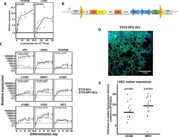
Fig. 6. Characterisation of (liver sinusoidal) endothelial cell markers in ETV2-ECs and ETV2-SPI1-ECs.
A ETV2-ECs were transduced on day 6 of differentiation with increasing titres of the SPI1-encoding lentiviral vector. The effect of SPI1 overexpression on the LSEC markers FCGR2B and LYVE1 is shown. Statistical analysis by linear modelling (expression ~ titre); N = 2 biological replicates per concentration. B ETV2-SPI1 construct engineered into the AAVS1 safe harbour locus. Recombination was performed analogously to Fig. 1A. (FRT Flippase recognition target, SA splice acceptor, PuroR puromycin resistance gene, TRE tetracycline response element, M2rtTA M2 reverse tetracycline transactivator). C Time-course comparison between ETV2-ECs and ETV2-SPI1-ECs for the overexpressed SPI1, for the endothelial cell-specific gene CDH5, for the known LSEC markers FCGR2B, LYVE1, MRC1, STAB1, and STAB2, and for novel LSEC markers, such as FCN3, and OIT3. Statistical significances were assessed by mixed ANOVA. Undetected mRNA values were represented with a relative expression of 1. The x-axes denote the days of differentiation. The differentiation protocols for ETV2-ECs and ETV2-SPI1-ECs are identical. D Tube formation assay on day 12 of differentiation of ETV2-SPI1-ECs. E LSEC marker expression (CD32B and MRC1) in ETV2-SPI1-ECs compared to ETV2-ECs. Chart indicates the median fluorescence intensity shift for CD32B and MRC1 of ETV2-ECs and ETV2-SPI1-ECs on day 12 of differentiation (replicates: N = 15 for CD32B and N = 14 for MRC1). The CD32 and MRC1 positive cells were gated from single-cell, PI-negative, and VE-cadherin-positive populations (gating strategy in Supplementary Fig. 3).

