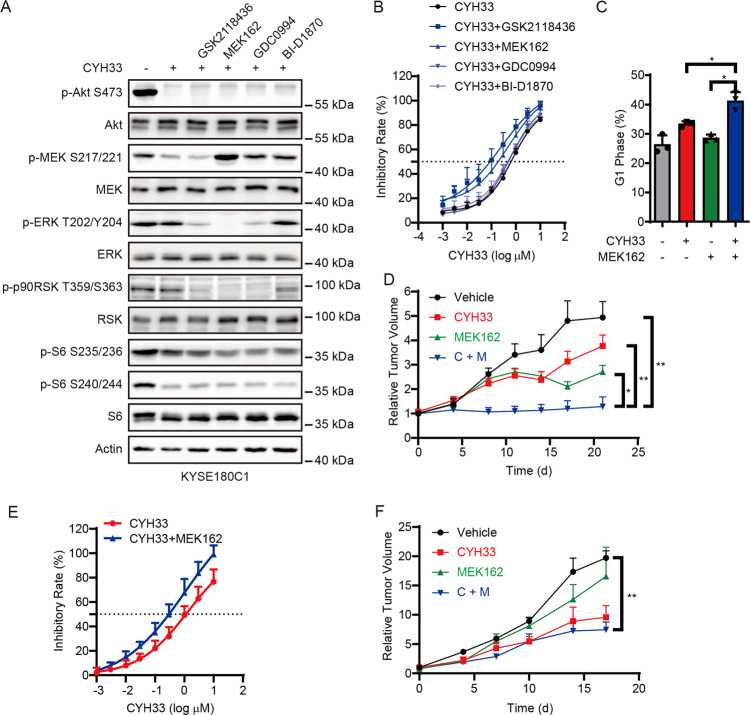
Fig. 5. Inhibition of MAPK signaling pathway sensitized KYSE180C cells to CYH33.
A KYSE180C1 cells were treated with 1 μM CYH33 alone or in combination with 1 μM GSK2118436/MEK162/GDC0994/BI-D1870 for 1 h. Cell lysates were subjected to western blotting with indicated antibodies. B KYSE180C1 cells were treated with serially diluted CYH33 alone or concurrently with 1 μM GSK2118436/MEK162/GDC0994/BI-D1870 for 72 h. Cell proliferation was measured with SRB assay. Data were presented as mean + SD (n = 3). C KYSE180C1 cells were treated with CYH33 (1 μM) and MEK162 (1 μM) alone or concurrently and cell cycle distribution was analyzed by flow cytometry. Percentage of cell population in G1 phase was presented as mean + SD (n = 3). Differences between the indicated groups were analyzed using one-way ANOVA with Tukey multiple group comparison test. *P < 0.05. D Randomly grouped nude mice bearing KYSE180C1 xenografts were administrated orally with a vehicle control, CYH33 (10 mg/kg), MEK162 (5 mg/kg), or a combination of CYH33 and MEK162 once a day for 21 days (n = 6). Tumor volume was measured twice a week. Data were presented as mean + SEM. Differences between the indicated groups were analyzed using one-way ANOVA with Tukey multiple group comparison test. **P < 0.01. E KYSE180-H cells were treated with serially diluted CYH33 alone or concurrently with 1 μM MEK162 for 72 h. Cell proliferation was measured with SRB assay. Data were presented as mean + SD (n = 3). F Randomly grouped nude mice bearing KYSE180-H xenografts were administrated orally with a vehicle control, CYH33 (10 mg/kg), MEK162 (5 mg/kg), or a combination of CYH33 and MEK162 once a day for 17 days (n = 3). Tumor volume was measured twice a week. Data were presented as mean + SEM. Differences between the indicated groups were analyzed using one-way ANOVA with Tukey multiple group comparison test. *P < 0.05.

