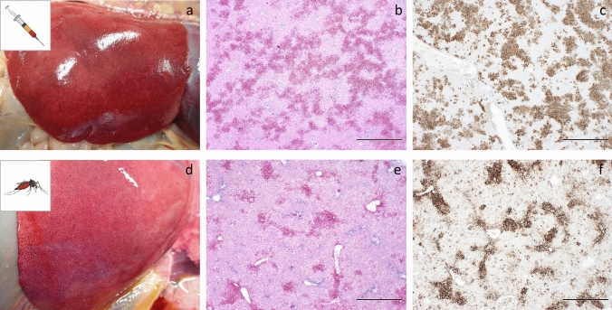
Figure 3.
Representative pathology in lambs exposed to RVFV via IV-inoculation or infected mosquitoes. Panels a–c were obtained from lamb #161 (IV inoculation). Panels d–f were obtained from lamb #154 (mosquito exposure). Both lambs were euthanized after reaching a HEP. Swollen liver with mottled appearance indicative of hepatic degeneration/necrosis (a,d), H&E staining of liver sections showing multifocal to bridging necrosis of hepatocytes with haemorrhages (b,e). IHC staining of a liver section with mAb 4-D4 showing strong immunolabelling for RVF antigen of the areas with degeneration and necrosis of hepatocytes (c,f). Bar = 1,000 μm.

