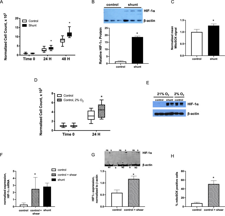
Figure 3.
Stabilization of HIF-1α is associated with faster LEC growth and accumulation of mitochondrially-derived reactive oxygen species (ROS). (A) shunt LECs proliferate with doubling time 15.2% faster than control LECs, *, P < 0.05, N = 3 control, 3 shunt, and (B) have 13.1-fold higher expression of HIF-1α, P < 0.05, N = 3 control, 3 shunt. NB, bar graph represents HIF-1α protein normalized to β-actin. (C) Mitochondrially-derived ROS were increased 1.3-fold in shunt LECs compared to controls, P < 0.05. N = 5 control, 5 shunt. NB, bars represent MitoSOX signal normalized to control. (D) control LECs cultured in 2% O2 proliferate significantly faster than control LECs cultured in 21% O2, *, P < 0.05; N = 3 control + 21% O2, 3 control + 2% O2. (E) HIF-1α is stabilized in control LECs cultured in 2% O2. (F–H) In control LECs exposed to 0.9 N/m2 of shear for 24hrs using a parallel plate flow chamber (ibidi Pump System), (F) expression of HIF-1α mRNA is significantly increased compared to levels in non-sheared control LECs, *, P < 0.05, N = 3 control no shear, 3 control + shear, 3 shunt; (G) similarly, HIF-1α protein is significantly stabilized in sheared (s) control LECs compared with non-sheared (ns) controls, *, P < 0.05; N = 3 control no shear, 3 control + shear; and, (H) 51% ± 8% sheared control LECs stained positive with MitoSOX compared with 8% ± 2% in non-sheared control LECs, *, P < 0.05; N = 3 control no shear, 3 control + shear. In panel F, blank lanes marked by ‘b’. In panels A, B, D, F, G, and H, error bars represent standard deviation. For panels B, E, and G, full-length blots/gels are presented in Supplemental Fig. 6.

