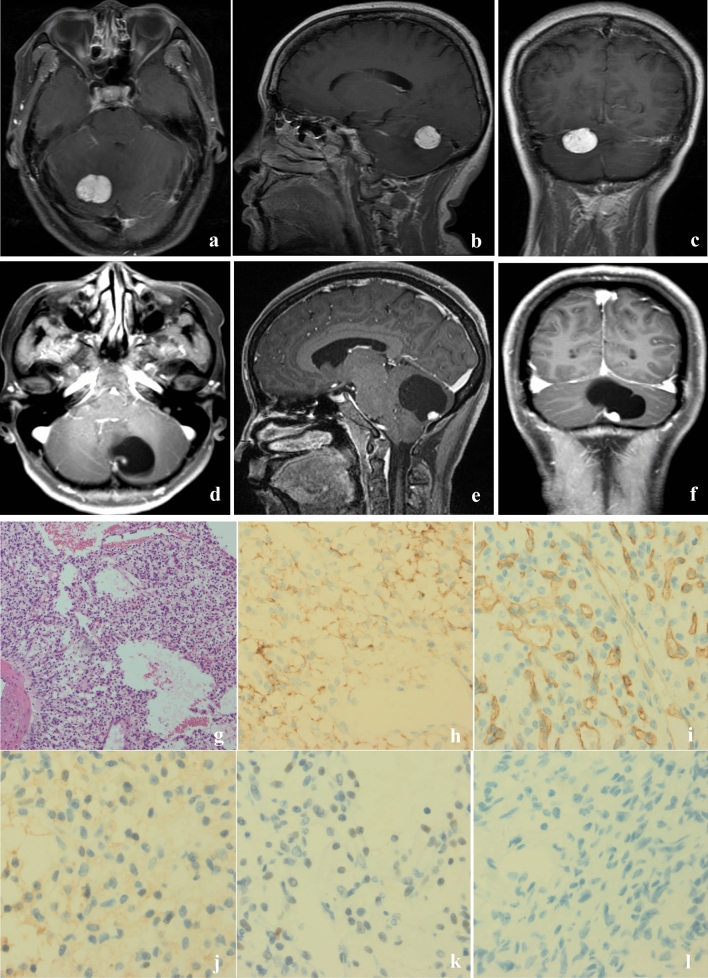
Figure 1.
Radiographic and immunohistochemical features of HBs. (a–c) Axial, sagittal and coronal contrast enhanced T1-weighted MRI of a solid HB showing well-circumscribed solid mass in the right cerebellar hemisphere. (d–f) Axial, sagittal and coronal contrast enhanced T1-weighted MRI of a cystic HB showing a non-enhanced cystic mass in the right cerebellar hemisphere with an enhancing mural nodule. The enhanced tumor nodule contained tumor cells, and surrounding cyst consisted of plasma ultrafiltrates. (g) HE staining demonstrated vascular and stromal cells in cystic HBs, ×100. Immunohistochemical staining of cystic HBs revealed tumor cells staining positive for carbonic anhydrase IX (CAIX) (h); endothelial cell marker (CD34) (i); 2-phospho-D-glycerate hydrolase (NSE) (j); SOX9 (k) and negative for phosphoenolpyruvate Carboxykinase (PCK) (l), ×400.

