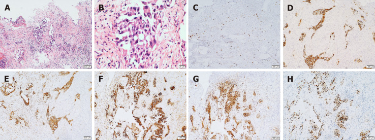
Figure 2.
Immunohistochemistry results. A: Increased nucleus and mitosis (hematoxylin and eosin × 10); B: Low differentiation and increased mucus secretion (positive hematoxylin and eosin × 40); C: Ki-67, suggestive of active cell proliferation and low degree of differentiation; D: Cytokeratin (CK)-positive, suggestive of epithelial cell differentiation, and thus, supporting the diagnosis of lung cancer metastasis; E: CK7 (+), supporting the diagnosis of lung cancer; F: CK-pan (+), suggesting that the tissue originated from epithelial cells; G: napsin A (+), suggestive of primary lung adenocarcinoma; H: thyroid transcription factor 1 (+), suggestive of non-small cell lung cancer.

