
Figure 1.
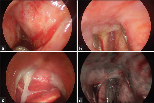
(a) A large portion of mucosa sloughing off in the epiglottic vallecula; (b) a magnified view of mucosal peeling in the anterior commissure region; (c) an endoscopic view of a large erythematous lesion involving the right vestibular fold; (d) view of mucosal peeling in the anterior commissure region with polarized light NBI to visualize the blood vessels