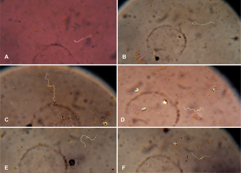
Fig. 3.
Microscopic observation of Leptospira with a 100× magnification optical microscope
A: Multiplying Leptospira; the image relates to pathogenic Leptospira spp., B: The Leptospira bacterium that is twisted over itself is located on the right. In the lower-left a shorter Leptospira is visible; the image related to Leptospira ryugenii, C and E: Long Leptospira, D: The hook area of the Leptospira bacterium was clearly observed in a microscopic view; the image related to Leptospira perdikensis, F: Two Leptospira bacteria that are interconnected from one end and form a long separable leptospiral chain; the image related to Leptospira sp.

