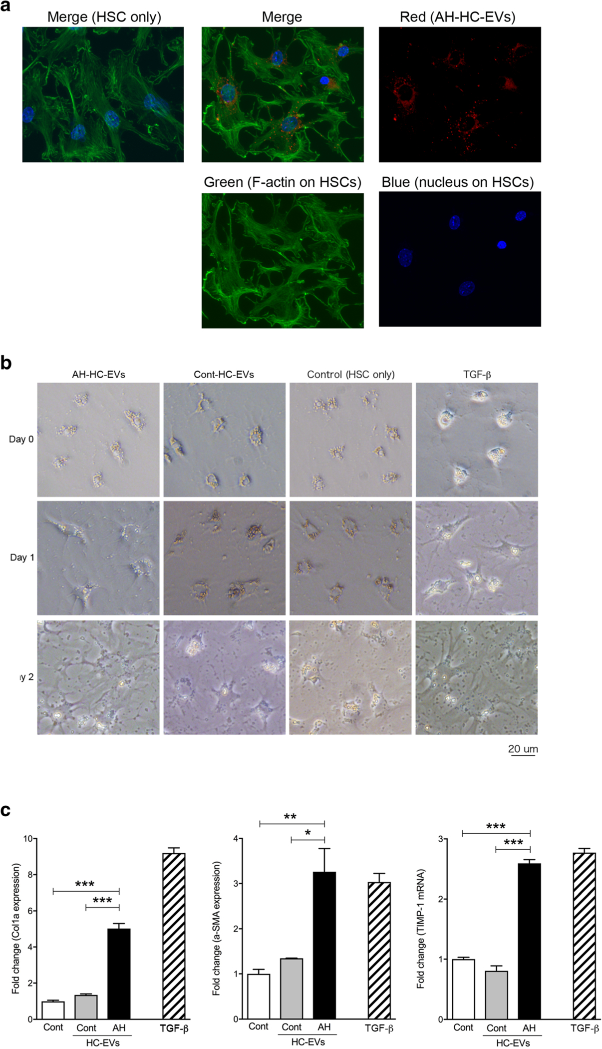
Fig. 3.

HC-EVs from AH activate primary HSC through miRNAs in HC-EVs. a Image of HSCs with labeled HC-EVs from AH mice. Green: FITC-phallotoxins and blue: DAPI in HSCs and red: HC-EVs with PKH26. b Morphology of primary HSCs with AH-HC-EVs, Cont-HC-EVs, control, and TGF-β1 at day 0, 1, and 2. c Fold change of Col1a, α-SMA, and TIMP-1 mRNA expression in primary HSCs with AH-HC-EVs, Cont-HC-EVs, control, and TGF-β at day 2. ***P < 0.001 and **P < 0.01. Values are mean ± SEM
