Fig. 5.

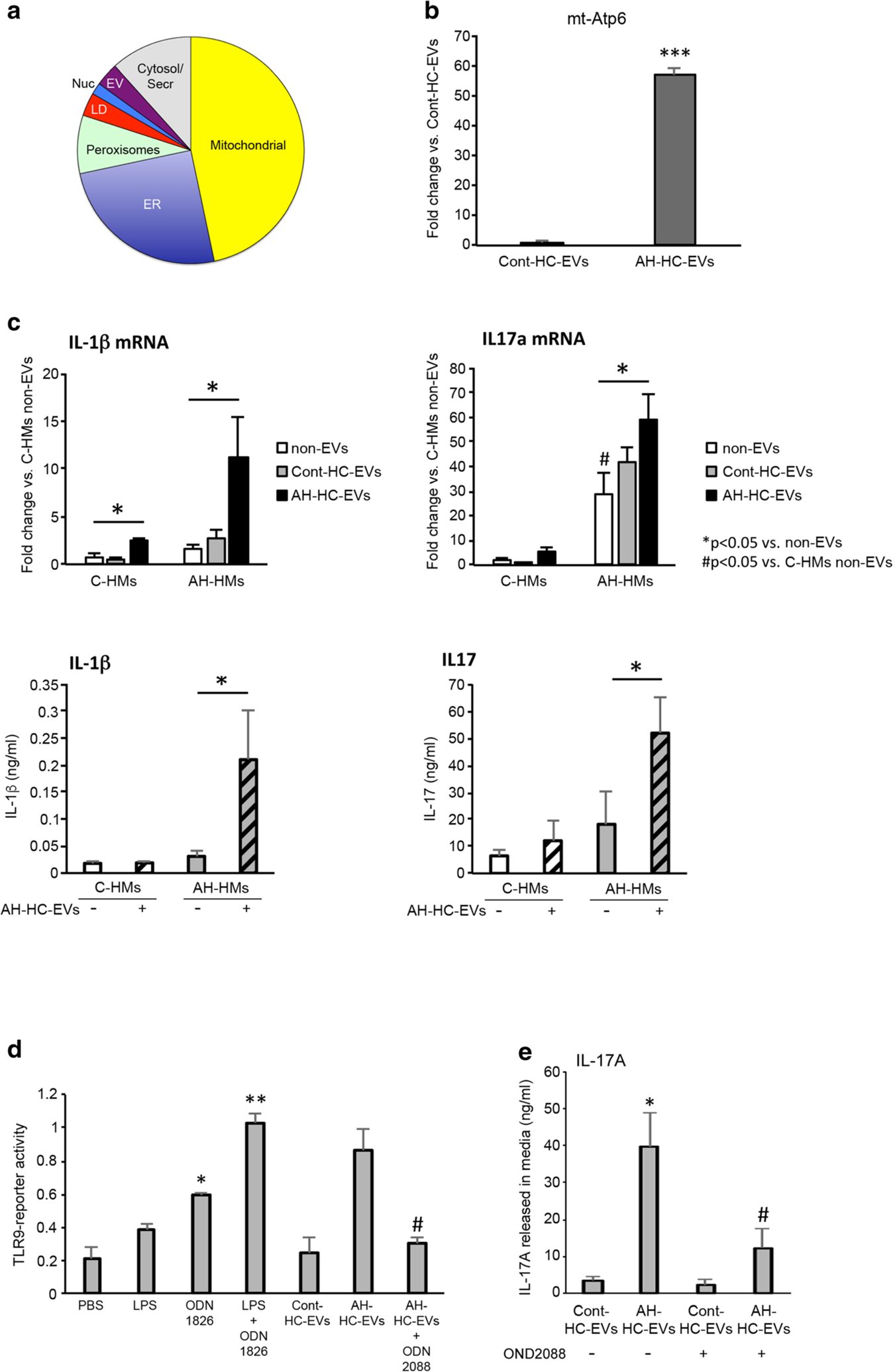
DAMPs in HC-EVs mediate hepatic macrophage activation through TL9 pathway. a Pie chart of upregulated protein composition in AH-HC-EVs (fold change >2). b Fold change of mitochondrial Atp6 (encoding the ATP synthase Fo subunit 6) DNA expression in AH-HC-EVs and Cont-HC-EVs. ***P < 0.001. c Fold change of IL-1β or IL-17a mRNA levels and protein production in hepatic macrophages (HMs) from pair-fed control or AH mice. *P < 0.05 vs. non-EVs. #P < 0.05 vs. C-HMs non-EVs. d TLR9 reporter activity in HEK-Blue-mTLR9 reporter cells incubated with LPS, ODN1826 (TLR9 agonist), LPS plus ODN1826 (TLR9 agonist), Cont-HC-EV, AH-HC-EVs, AH-HC-EVs plus ODN2088 (TLR9 antagonist). **P < 0.01 and *P < 0.05 vs. PBS. #P < 0.05 vs. AH-HC-EVs alone. e IL17A production in supernatant of AH-HMs incubated with Cont-HC-EVs and AH-HC-EVs without or with ODN2088. *P < 0.05 vs. Cont-HC-EVs. #P < 0.05 vs. AH-HC-EVs alone. Values are mean ± SEM. C-HMs, control HMs; ER, endoplasmic reticulum; LD, lipid droplets; EV, extracellular vesicle; Serc, secretary compartment
