
Figure 1.
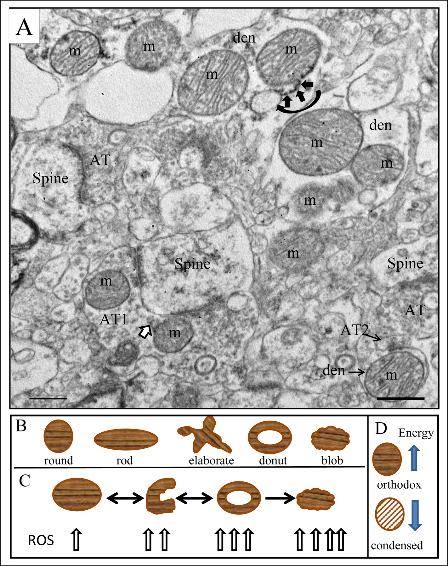
A) Electron micrograph of human striatum. Mitochondria (m) are indicated in various subcellular locations. In the dendrite (den) at the top of the field, a mitochondrial associated ER (MAM) is shown (curved black arrow) with ER (short black arrows) connecting to the adjacent mitochondrion. Axon terminal (AT1) forms an excitatory synapse on a spine in the lower part of the field; mitochondrial derived vesicles (MDVs) are shown (white arrow with black outline) budding off of a mitochondrion in the terminal. Axon terminal AT2 forms an inhibitory synapse on the dendrite (den). Scale bar = 0.5 µm. Figure is modified from Figure 2a in Somerville et al., 2011b and Figure 1 in Roberts, 2017). B) Drawings of different shaped mitochondria. C) Transformation of a mitochondrion from round/rod to curved to donut to blob shape and the corresponding amount of reactive oxygen species each produces. Arrows are bidirectional between round and curved and donut shaped mitochondria indicating the ability to change shape in either direction. Once a mitochondrion has assumed a blob shape, it cannot recover healthier configurations, thus the unidirectional arrow. D) Depiction of the orthodox and condensed form of mitochondria. Orthodox configuration is high energy producing, while condensed configuration is low energy producing, indicated by the directionality of the arrows.