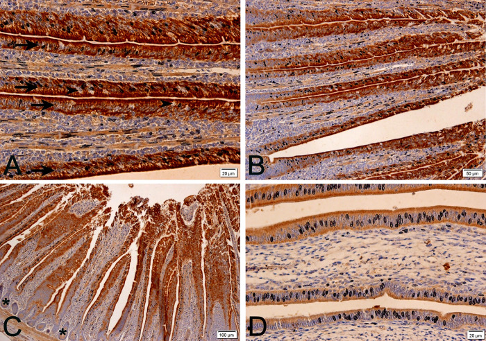
Fig 1. Photomicrographs of anti-Calbindin-D28k immunohistochemistry in laying quail intestine at different magnifications.
Positive (brown stain) anti-calbindin-D28k intestinal epithelium (arrows) and non-positive goblet cells (arrowheads) (A and B) are observed. Non-antibody-positive crypts (asterisk) are also observed (C). Lower epithelial positivity (brown stain) is observed under 28°C (D) when compared to other temperature treatments. Chromogen staining diaminobenzidine+hematoxylin.

