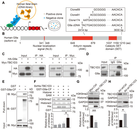
Fig. 2. Interaction between TBC1D3 and G9a inhibits H3K9me2 modification.

(A) Schematic diagram for Y2H screening assay. (B) Domain structure of human G9a protein and sequences of positive clones. (C) IB analysis of reciprocal co-immunoprecipitation (co-IP) results in HEK293T cells transfected with HA-G9a or HA-G9a plus Myc-TBC1D3. (D) Homogenates of GW15 human fetal cortical tissues were subjected to IP with anti-G9a antibody with immunoglobulin G (IgG) as a control and IB with antibodies against TBC1D3 or G9a. Shown is an example of two independent experiments with similar results. (E) Homogenates of HEK293cells transfected with Myc-TBC1D3 were subjected to pull-down with beads coupled with GST or GST-G9a-CF [649 to 1210 amino acids (aa)], followed by IB with anti-Myc antibody. (F) Addition of 6xHis-TBC1D3 attenuates the Histone3 methylation activity of G9a. Relative levels of H3K9me2 with respect to that of Histone3 from three independent experiments were quantified. (G) Levels of H3K9me2 in ReN cells transfected with control (Myc-Ctrl) or Myc-TBC1D3 plasmid (six independent experiments). (H) Levels of H3K9me2 in human cerebral organoids infected with AV-shCtrl or AV-shTBC1D3 (three independent experiments). The quantified data are presented as means ± SD by unpaired Student’s t test. *P < 0.05; **P < 0.01. bp, base pair.
