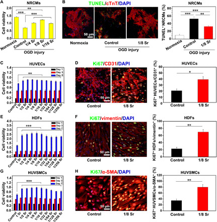
Fig. 1. Sr ions reduce NRCM apoptosis after OGD injury and promote blood vessel–related cell proliferation.

(A) The viability of NRCMs measured by Cell Counting Kit-8 (CCK8) in the medium supplemented with different concentrations of Sr ion after OGD injury. (B) TUNEL staining (green), cTnT staining (red), and DAPI (4′,6-diamidino-2-phenylindole) staining (blue) in NRCMs after OGD injury and quantitative analysis of TUNEL+ NRCMs (10 pictures for each group). The corresponding concentrations of Sr ion with the 1/4 to 1/16 dilution ratio for NRCM culture are shown in table S1. (C to H) The cell viability and proliferation of human umbilical vein endothelial cells (HUVECs) (C and D), human dermal fibroblasts (HDFs) (E and F), and human umbilical vein smooth muscle cells (HUVSMCs) (G and H) after culturing in the medium supplemented with Sr ions at different concentrations. They were respectively revealed by CCK8 and the immunofluorescence of Ki67, followed by quantitative analysis of Ki67+ after Sr ion treatment for 5 days (HUVECs) or 7 days (HDFs and HUVSMCs). The corresponding concentrations of Sr ion with the dilution ratio of 1 to 1/256 for HUVEC, HDF, and HUVSMC culture are respectively shown in table S1. Experiments were conducted in triplicate. All data are presented as means ± SEM. An unpaired t test was used to compare between any two groups. One-way analysis of variance (ANOVA) was used to compare between three or more groups. *P < 0.05, **P < 0.01, and ***P < 0.001.
