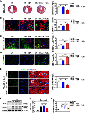
Fig. 6. SrCO3/HSA hydrogels inhibit myocardial fibrosis, increase angiogenesis, and reduce cardiomyocyte apoptosis.

(A) Representative cross-sectional images and quantitative data of scar area in LV on 5-μm slices stained with Masson’s trichrome at day 28 after I/R. n = 5 hearts for each group. (B to D) Images and quantification of α-SMA+ blood vessels (B), CD31+ cells (C), and VEGF (D) in the border zone of infarcted hearts 28 days after I/R. Scale bars, 50 μm. n = 4 hearts for each group. HPF, high-power field. (E) Representative and quantification of staining for TUNEL+ cardiomyocytes (CMs) in the border zone of infarcted hearts at day 3 after I/R. Scale bars, 50 μm. n = 5 to 6 hearts for each group. (F and G) Representative and averaged Western blot analysis for cleaved caspase-3 (cCas.3) and glyceraldehyde 3-phosphate dehydrogenase (GAPDH) in LV heart tissues at day 3 after I/R. n = 4 hearts each. (H) The concentrations of cardiac injury marker LDH of the mice serum at day 3 after I/R. n = 4 to 6 hearts each. Ctrl, I/R control; HSA, I/R + HSA; 1% Sr, I/R + HSA + 1% Sr. All values are expressed as means ± SEM; one-way ANOVA was used for statistical analyses. *P < 0.05 and ***P < 0.001.
