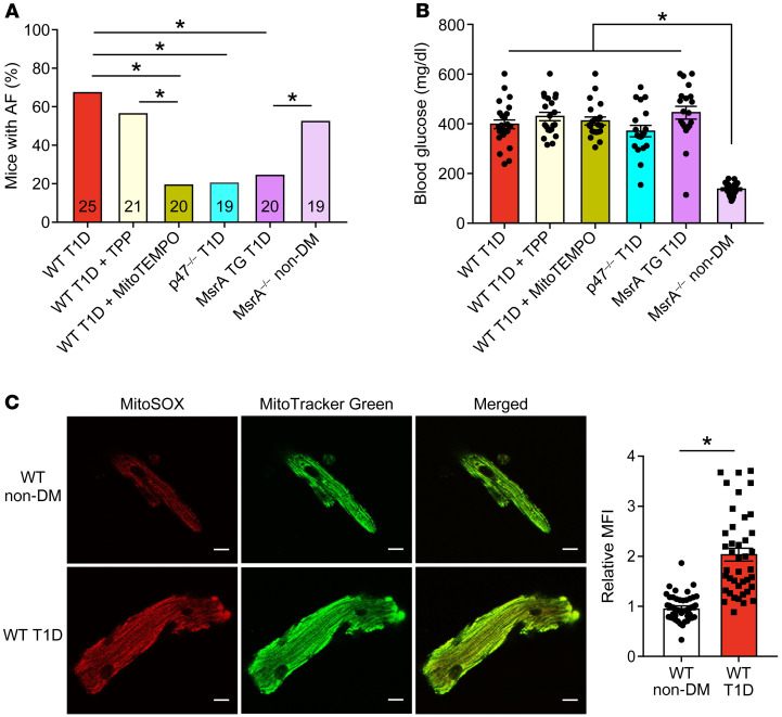
Figure 5. Targeted ROS inhibition and MsrA overexpression protects against atrial fibrillation in diabetes.
(A) Inhibition of mitochondrial ROS by MitoTEMPO treatment or inhibition of cytoplasmic ROS by loss of the p47 subunit of NADPH oxidase (p47–/– mice) protect against AF in T1D mice. Mice with myocardial targeted transgenic overexpression of methionine sulfoxide reductase A (MsrA TG) were protected from T1D primed AF; nondiabetic mice lacking MsrA (MrsA–/– non-DM) showed increased AF susceptibility in the absence of diabetes. (B) Summary data of blood glucose measurements at the time of electrophysiology study. (C) Increased mitochondrial ROS in isolated atrial myocytes from WT T1D (bottom) compared with WT non-DM (top) mice detected by MitoSOX fluorescence. Representative confocal fluorescent images (original magnification, ×40) show MitoSOX (red, left), MitoTracker (green, middle), and merged images (right). Scale bars: 10 μm. (n = 41–47 cells in each group from 2 mice per group). Data are represented as percentage frequency distribution (A) and mean ± SEM (B and C). The numerals in the bars represent the sample size in each group (A). Statistical comparisons were performed using 2-tailed Fischer’s exact test with Holm-Bonferroni correction for multiple comparisons (A), 1-way ANOVA with Tukey’s multiple-comparison test (B) and 2-tailed Student’s t test (C). (*P < 0.05). WT T1D data set (A and B), control data previously presented. AF, atrial fibrillation; DM, diabetes mellitus; T1D, type 1 DM; T2D, type 2 DM.

